Professor Majlinda Lako from Newcastle University reflects on her career for International Women's Day 2026
I created my own research group in Newcastle in 2003. At the time, the focus was to set up a pluripotent embryonic stem cell research group. These were human embryonic stem cells derived from spare IVF embryos.
With a couple of colleagues, we were very successful. We deposited one of the first two UK human embryonic stem cell lines in the UK stem cell bank.
But even then, I was already building collaborations with ophthalmic clinicians.
Developing an attraction for retinal research
At first, I wasn’t working in the retina. I was working in the front of the eye, the cornea. Within three years, we started clinical trials in Newcastle, bringing sight to people who had damage in their eyes — from fireworks, from accidents, from thermal or chemical burns.
Working closely with clinicians changed everything. They kept coming back to me and saying, 'You’ve got the stem cell expertise.' We’ve got the ideas. The retina — the back of the eye — is much more interesting. It’s much more complex.
A Nobel Prize breakthrough that accelerated progress
Around 2006, I began applying for funding to generate retinal models in the lab. It was lucky timing. Induced pluripotent stem cells had just been discovered — a Nobel Prize-winning breakthrough that allowed us to take adult cells and bring them back to an early, stem-cell-like stage.
We had human embryonic stem cells and induced pluripotent stem cells side by side. That’s when we started making retinal models.
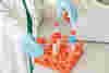
They are little organoids in the lab, but they have all the cells of the retina, and we could prove maybe in early 2015, 2016, they’re also light responsive, mimicking the main function of the retina.
That was a eureka moment.
I clearly remember thinking that all the sleepless nights, all the family sacrifices, had been worth it. We had shown that these lab-grown structures didn’t just look like retina. They behaved like it.
Expanding research into eye disease modelling
After that, the group became really established. I received a European Research Council Fellowship from 2013 to 2018 to develop the model in detail. We expanded into disease modelling — retinitis pigmentosa, age-related macular degeneration. We started transplanting cells in animal models. We looked at cancers like retinoblastoma and began drug repurposing.
Our research now incorporates understanding how disease arises, understanding how the retina forms during embryonic development, modelling disease, repurposing drugs and developing cell-based transplants.
But science always moves forward.
The next-generation of retinal organoids
Once we make retinal organoids, they grow and grow beautifully but comes a point where they grow too much.
Because they don’t have blood vessels, a dark, necrotic centre can form. Nutrients and oxygen can’t diffuse properly. Important cells begin to suffer.
That’s why we began working on what I call the next-generation organoids.
We and others are working on what we call next-generation organoids, which contain both blood vessels and immune cells.
If you are modelling diseases like diabetic retinopathy, you really need blood vessels there. You need immune cells. You need complexity.
It’s a more realistic disease modelling. To have the new generation organoid with vascular cells and immune cells compared to the one that was before, which didn’t have those.
We are also working on reproducibility. Even the same operator can generate different results from day to day. We need robotics. We need standardisation. We need scale.
To go to the clinic, we must be precise.
Eureka moments in the lab and beyond
There have been other moments that stay with me.
One was when we transplanted photoreceptors from retinal organoids into blind mice. We used a simple dark-and-light box test. Mice prefer the dark. The mice without transplants wandered randomly. But the transplanted mice spent most of their time in the dark.
Although their vision wasn’t perfect, they knew how to go to the dark and stay there.
That was another eureka moment.
I also feel proud of the retinoblastoma iPSC model we created. We’ve now identified two promising drug candidates that we want to optimise towards clinical trials. For the first time, I can really see the end goal.
Future possibilities in gene editing
And looking ahead, I am excited about gene editing.
I am excited about the progress made in gene editing. So CRISPR Cas and prime editing, I think they’re fantastic tools because, there is nothing better than being, if you have a mutation, being able to reverse that mutation early on so your cells don’t die and they function.
But I’m also realistic. Personalised therapies can be expensive. That’s why we are working on gene-agnostic approaches — creating photoreceptors that are invisible to the immune system, so they can be transplanted without rejection.
If we can freeze them and have them ready, then when a patient comes, we can act immediately.
On the cusp of future breakthroughs
I’ve lived through enormous scientific progress in my career. From the early days of induced pluripotent stem cells in 2007 to where we are now, the field has exploded.
“I think we are on the cusp.”
There are already gene therapy and cell transplant trials underway. If they prove safe and effective, there is nothing to stop larger trials moving forward in the next five to ten years.
I’ve seen how long research can take. I’ve seen how difficult it can be. But I’ve also seen breakthroughs.
If I am lucky enough to work another 20 or 30 years, I believe I will see even more progress.
And that keeps me going.